No Cell Left Behind®
EPIC SCIENCES TECHNOLOGY OVERVIEW
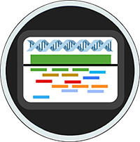
For hundreds of years, scientists have studied the cell to understand biological function and disease progression. The Epic Sciences team took these basic studies in cell morphology and intracellular biomarker localization one step further. We have developed a platform that enables you to identify and analyze rare disease cells from a tube of blood that also contains millions of normal cells. It’s a revolutionary digital pathology technology that acquires high-definition images of each cancer cell found in a sample that empowers characterization at single cell resolution.
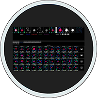
The Epic Sciences platform not only assesses the number of CTCs and CTC subtypes, but also profiles single cell phenotype and genotype:
- Phenotypic Measures
- Specific biomarker expression levels
- Subcellular biomarker localization
- Morphologic characteristics
- Genotypic Measures
- NGS
- FISH


UP TO DATE WITH EPIC SCIENCES
Check out our 2015 webinar entitled Real-Time Profiling of Tumor Heterogeneity in Metastatic Disease in which we review our platform.